agenesis of ICA(内頚動脈無形成症)
 |
 |
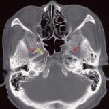 |
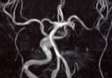
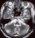
| MR Angio | MRI T2WI | CT |
|
|
|
|
| MR Angio | MRI T2WI | CT |
【図の説明】
症例は90歳女性。
MRアンギオでは左の内頚動脈が描出されていない。つい、内頚動脈閉塞症と言いたくなる所見である。ところが、MRI
T2強調画像を見ると左の内頚動脈の flow void のみならず、血栓からの信号も見えない。CTの骨条件では左側で卵円孔の背側にあるべき頚動脈管が形成されていないことがわかり、内頚動脈無形成(片側第1型)の診断が確定する。
【疾患の解説】
内頚動脈の無形成は内頚動脈の閉塞と病態は似ているが、頭蓋骨の頚動脈管(carotid
canal)も無形成〜低形成を示すことが異なる(鑑別診断のポイント)。
片側性のものと両側性のものとで血行動態が異なる。Lie は3型に分類している。
片側性では、
・第1型:ACA
は対側から、MCA は Pcom からの血流を受ける
・第2型:ACA と MCA はともに対側の
ACA から血流を受ける
・第3型:transsellar intercavernous intercarotid
anastomosis のある例
両側性では、さまざまな側副血行路が見られる。